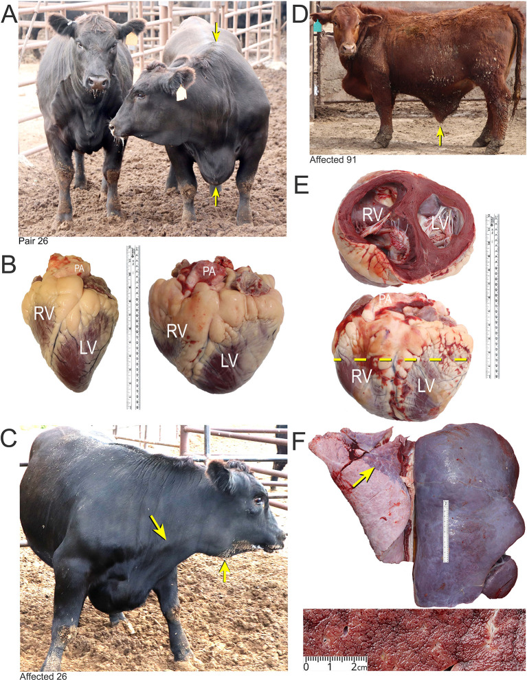
Figure 1. Features of BCHF in feedlot cattle.
Panel A, matched pair number 26 with the clinical case (right) showing ventral edema and a drop in the withers (arrows). The latter occurs as the fluid accumulates in thorax pushing the shoulders outward. Panel B, the affected heart of case 26 (right) compared with a normal heart of a fattened American Angus heifer. Abbreviations: RV, right ventricle; LV, left ventricle; PA, pulmonary artery. Panel C, PA distension and mandibular edema in case 26 (arrows). Panel D and E, clinical case number 91 with ascites (arrow) and enlarged heart with dilated RV. Panel F, Enlarged, encapsulated liver of case 91 with “nutmeg liver” appearance in cross section due to hepatic venous congestion, and atelectasis in lungs (arrow).

