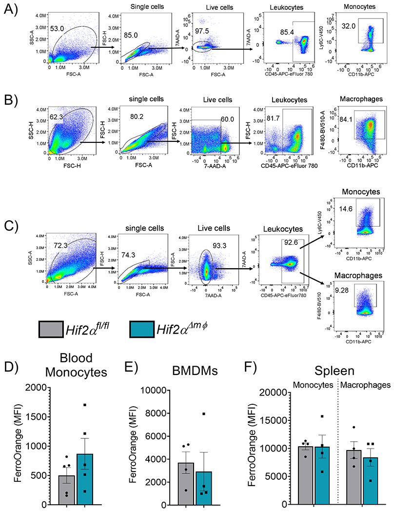
Figure 2. Monocyte differentiation and intracellular labile iron are not altered following HIF2α in myeloid cells.

Representative Gating strategy for (A) circulating blood monocytes (n=5,each group) (B) bone marrow derived macrophages (BMDMs) (n=4, each group) and (C) splenic monocytes (n=4, each group) or macrophages (n=4, each group). Mean fluorescence intensity of FerroOrange gated on (D) blood monocytes. (E) BMDMs or (F) splenic monocytes and macrophages. Data represent the mean ± SEM. Significance was determined by unpaired t test. *P < 0.05, ***P < 0.001, and ****P < 0.0001 versus the Hif2αfl/fl group.
