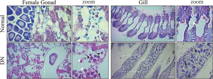
Fig. 1.
Disseminated neoplasia in Mytilus chilensis. To determine if MchSLEs are correlated with the presence of disseminated neoplasia in M. chilensis, individuals from 2 different farm sites were diagnosed. Animal were fixed and sliced before hematoxylin-eosin staining. The diagnostic considered level of hemocyte infiltration in the tissue, morphological characteristics, and nuclear size of infiltrating cells. Representative image of female gonad and gills of a normal and a disease animal are shown. A zoom of the area depicted by the withe square is also shwon. Withe arrow heads indicate normal circulating cells, while black arrows indicate cancer cells. DN: disseminated neoplasia. Size bar = 20 μm

