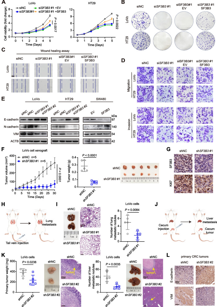
Fig. 2.
SF3B3 promotes the proliferation and metastasis of CRC in Vitro and Vivo. A Growth curves and (B) colony formation of LoVo and HT29 cells after SF3B3 knockdown by siRNAs (siNC vs siSF3B3), or after SF3B3 knockdown by siRNAs for 12 h followed with re-expression of SF3B3 (siSF3B3 + EV vs siSF3B3 + SF3B3) (empty vector, EV; SF3B3 overexpressing plasmid, SF3B3). Cell viability was determined at different time points and colony formation assay was measured after 2 weeks. C Wound healing assays and (D) Transwell assays of LoVo and HT29 cells to investigate the effects of SF3B3 knockdown (siNC vs siSF3B3) or re-expression (siSF3B3 + EV vs siSF3B3 + SF3B3) on cell migration and invasion abilities. Scale bars, 100 μm. E Representative western blots of EMT-related proteins in SF3B3-knockdown or SF3B3-overexpressing CRC cells. F Tumor growth, tumor weights, and tumor images of xenografts in nude mice. LoVo cells were infected with shNC or shSF3B3#1 lentivirus to obtain the stably cell clones, which were subcutaneously injected into flank region of each nude mouse. G Representative IHC images of SF3B3 and Ki67 proteins in xenografts. Scale bars, 25 μm. H Schematic design of the CRC lung metastasis mouse model. I Representative images of lung, H&E staining of lung tissues derived from mice after tail vein injection with stably SF3B3-knockdown LoVo cells (LoVo-shSF3B3#1), and statistical analysis of lung metastatic nodules. Scale bars, 200 μm. J Schematic design of the CRC liver metastasis mouse model. K Statistical analysis of primary CRC tumor weights and liver metastatic nodules, as well as representative H&E staining. Stably SF3B3-knockdown LoVo cells were constructed using shSF3B3#2 lentivirus. Scale bars, 200 μm. L Representative IHC images of E-cadherin and VIM in primary CRC tumors from mice orthotopically injected with SF3B3-knockdown LoVo cells (LoVo-shSF3B3#2). Scale bars, 100 μm. Data are presented as mean ± SD

