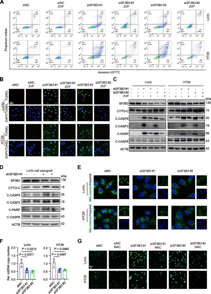
Fig. 3.
Silencing of SF3B3 induces the intrinsic apoptotic pathway in CRC cells. A Detection of apoptosis by Annexin VFITC/PI staining and flow cytometric analysis. LoVo and HT29 cells were transfected with siRNAs for 48 h followed by 20 μM ZVF (Z-VAD-FMK) treatment for 24 h. B Representative images of TUNEL staining. Nuclei (blue) were stained with DAPI. CRC cells were transfected with siRNAs for 48 h followed by 20 μM ZVF treatment for 24 h. Scale bars, 50 μm. C Representative western blots of apoptosis-related proteins in CRC cells. LoVo and HT29 cells were transfected with siRNAs for 48 h, followed by 20 μM ZVF treatment for 24 h. D Representative western blots of apoptosis-related proteins in LoVo xenografts (shNC vs shSF3B3#1). E Representative fluorescence images of MitoTracker (green) staining using a confocal microscope with 63 × oil immersion lens. Nuclei (blue) were stained with Hoechst 33342. LoVo and HT29 cells were treated with siRNAs for 72 h. F mtDNA copy number was quantified by qRT-PCR. LoVo and HT29 cells were treated with siRNAs for 48 h. Data are shown as mean ± SD. G Representative fluorescence images of ROS (green) staining. LoVo and HT29 cells transfected with siRNAs for 24 h, followed by 10 mM NAC (N-acetylcysteine) treatment for 48 h. Scale bars, 50 μm

