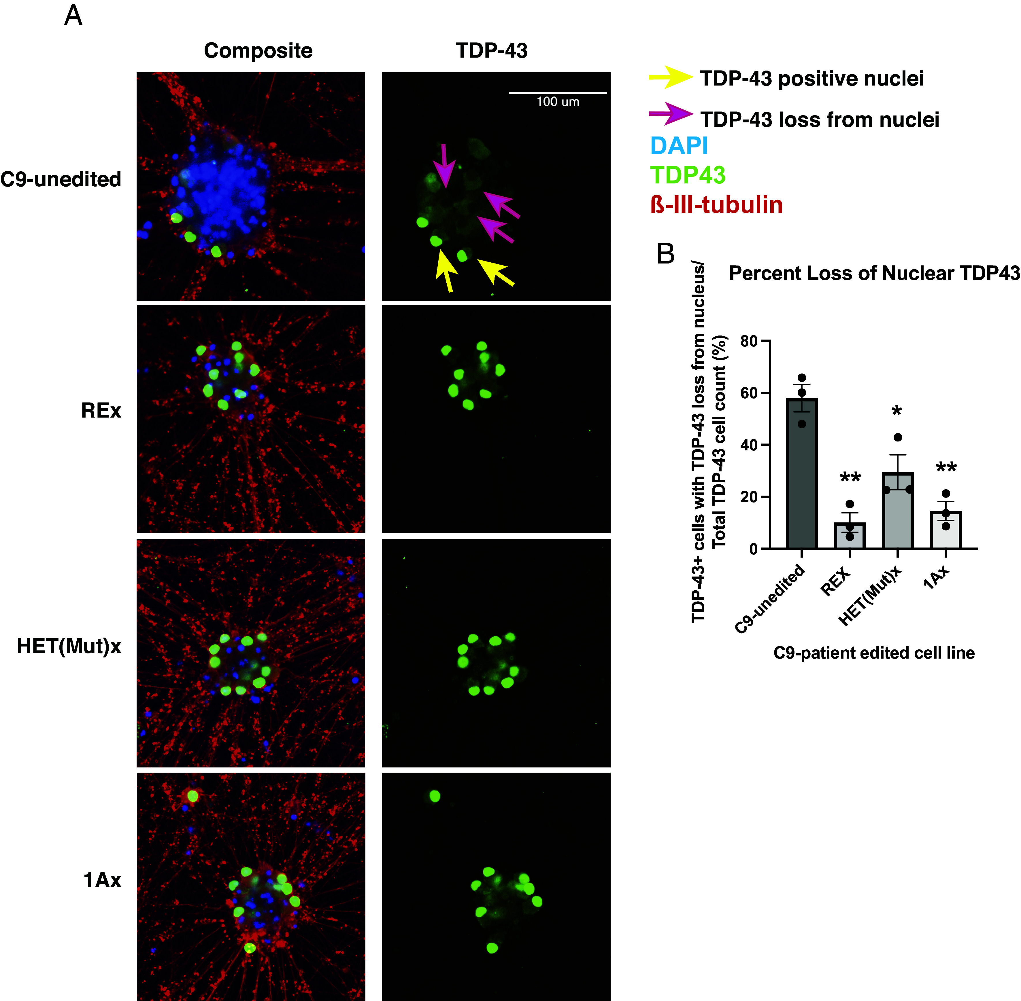
Fig. 4.

Three editing approaches ameliorate loss of nuclear TDP-43 in 7-wk-old neurons derived from C9 iPSC line. (A) Immunofluorescent images of neurons derived from unedited and edited C9 iPSCs. The neurons were grown for 7 wk and stained for TDP-43 (green), DAPI (blue) and beta-III-tubulin (red). Yellow arrow points to an example nuclei harboring TDP-43 and pink arrows to example TDP-43-positive cells whose nucleus are devoid of TDP-43. (B) Percentage of TDP-43-positive cells that lack nuclear TDP-43 (1-way ANOVA F(3,8) = 18.65; P < 0.001; *P < 0.05, **P < 0.01 by Tukey’s multiple comparison’s post hoc test between C9-unedited and each sample. REx, HET(Mut)x, and 1Ax were not statistically significantly different from one another). Each experiment contained three biologic replicates (separate wells). Error bars = SEM.
