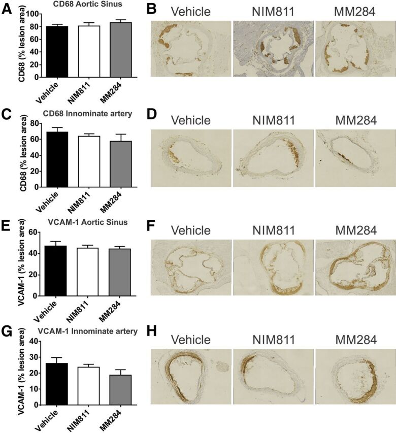
Fig. 2.

Cyclophilin inhibition and markers of inflammation in the atherosclerotic plaque. (A) Quantitation of CD68 staining for macrophages in the aortic sinus. Percentages of anti-CD68–stained area of total plaque area are shown. (B) Representative micrographs of the CD68 staining in the aortic sinus of mice treated with vehicle (left), NIM811 (center), and MM284 (right). (C) Quantitation of CD68 staining for macrophages in the innominate artery. (D) Representative micrographs of the innominate artery stained for CD68 of mice treated with vehicle (left), NIM811 (center), and MM284 (right). (E) Quantitation of VCAM-1 content of lesions in the aortic sinus. Percentages of anti-VCAM-1–stained area of total plaque area are shown. (F) Representative micrographs of VCAM-1 staining in the aortic sinus of mice treated with vehicle (left), NIM811 (center), and MM284 (right). (G) Quantitation of VCAM-1 content of lesions in the innominate artery. Percentages of anti-VCAM-1–stained area of total plaque area are shown. (H) Representative micrographs of the VCAM-1 staining in the innominate artery of mice treated with vehicle (left), NIM811 (center), and MM284 (right). All graphs are presented as mean ± S.E.M.
