Fig. 4.

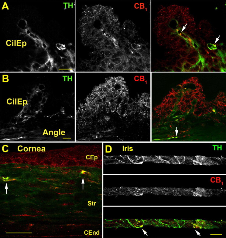
CB1 colocalizes with TH in the murine anterior eye. A, TH (left) and CB1 staining (center) in the ciliary epithelium (CilEp), and TH (green) and CB1 (red), with overlapping staining in yellow (arrows) (right). B, overview of ciliary epithelium (CilEp); including the angle shows TH and CB1 staining in the angle (arrow). C, in the cornea, CB1 is present in corneal epithelium (CEP), stroma (Str), and corneal endothelium (CEnd) and colocalizes with TH in the peripheral distal stroma (arrows). D, CB1 exhibits partial overlap with TH in the iris (arrows). Scale bars, 25 μm.
