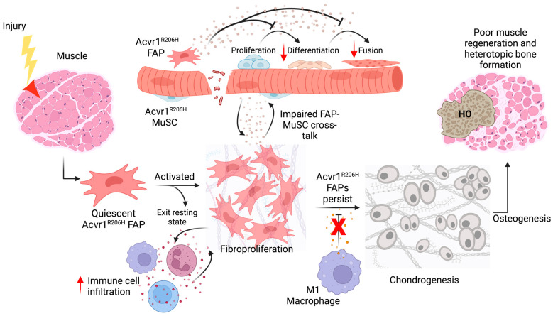
Figure 2.
Model for FOP muscle regeneration. After muscle injury, quiescent ACVR1R206H FAPs become activated and proliferate. However, abnormal FAP-derived soluble secretions decrease ACVR1R206H MuSC myogenic commitment and ability to fuse to pre-existing myofibers. At the same time, there is increased immune cell infiltration, while FAPs resist macrophage-derived TNFa-mediated apoptosis and continue to accumulate within the FOP tissue, giving rise to aberrant osteochondrogenesis. This leads to reduced muscle regeneration and increased heterotopic ossification in FOP tissue. Created with BioRender.com.

