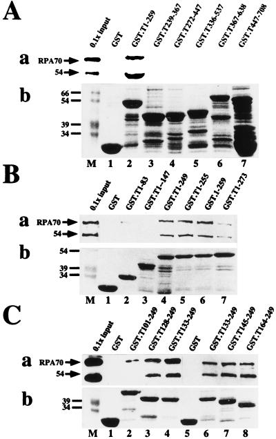
FIG. 1.
Mapping the RPA binding sequences of SV40 T antigen. The indicated residues of T antigen were expressed as GST fusion proteins and adsorbed to glutathione-agarose. Fusion protein-bound beads were incubated with purified RPA in a pull-down assay. (a) After washing, bound RPA was detected by denaturing gel electrophoresis and immunoblotting with RPA antibody 70C and chemiluminescence (lanes 1 to 7 [A and B] or 1 to 8 [C]). As a marker, 1/10 of the input RPA (lanes M) was analyzed in parallel. Positions of the 70-kDa subunit and a 54-kDa degradation product are indicated by arrows. (b) A 10-μl sample of beads bearing each fusion protein was analyzed by denaturing gel electrophoresis and detected by Coomassie staining (lanes 1 to 7 and 1 to 8). Lanes M show prestained marker proteins. Only the relevant portions of the gels are shown.

