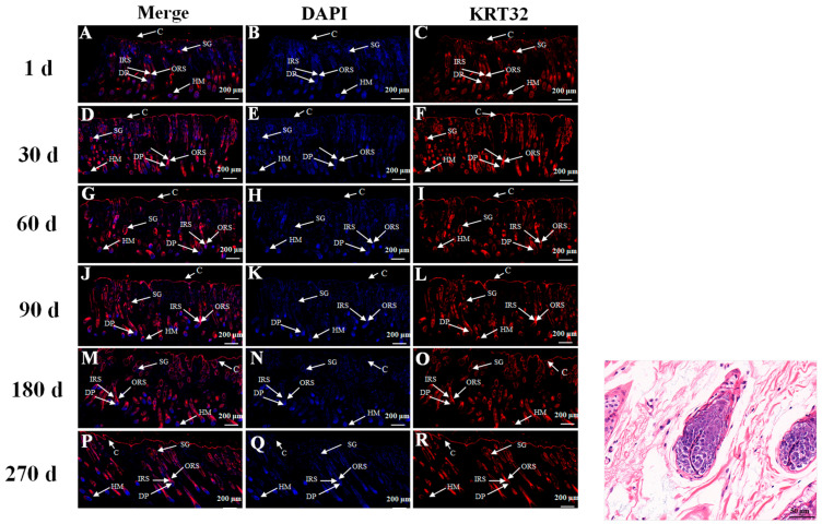
Figure 2.
Immunofluorescence staining for K32 in skin tissues of Gansu Alpine Fine-wool sheep at different times (5×). Blue (B,E,H,K,N,Q) and red (C,F,I,L,O,R) tissue in the figure show DAPI-labeled nuclear fluorescence staining and fluorescence staining for K32, respectively. ORS: outer root sheath; IRS: inner root sheath; HM: hair matrix; C: corneum; SG: sebaceous glands; DP: dermal papilla. H&E staining image are shown on the right. The first column (A,D,G,J,M,P) is a combined graph of the second and third columns.

