Figure 3.
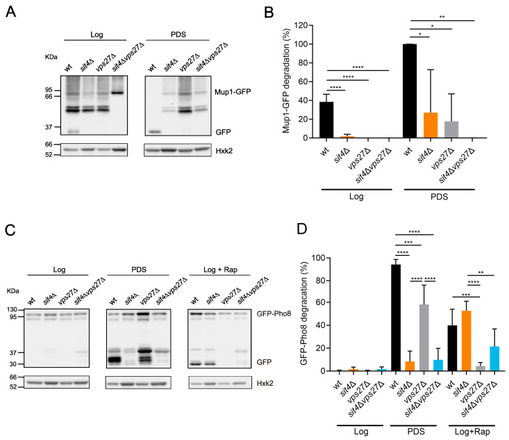
The induction of the MVB pathway and microautophagy at the PDS phase is Sit4-dependent. (A) Immunodetection of Mup1-GFP and GFP in protein extracts from cells expressing pRS416-MUP1-GFP grown in SC medium. Hxk2 was used as a loading control. A representative blot is shown. (B) The induction of the MVB pathway (free GFP/(Mup1-GFP + GFP) ratio) was evaluated at the Log and PDS (24 h after Log) phases. Values are the mean ± SD (n = 3); * p < 0.05, ** p < 0.01, **** p < 0.0001; one-way ANOVA. (C) Immunodetection of GFP-Pho8 and GFP in protein extracts from cells expressing pRS426-GFP-PHO8. Hxk2 was used as a loading control. A representative blot is shown. (D) The induction of microautophagy (free GFP/(GFP + GFP-Pho8) ratio) was evaluated at the Log and PDS (24 h after Log) phases. Cells treated with rapamycin were used as a positive control. Values are the mean ± SD (n = 3); ** p < 0.01, *** p < 0.001, **** p < 0.0001; one-way ANOVA.

