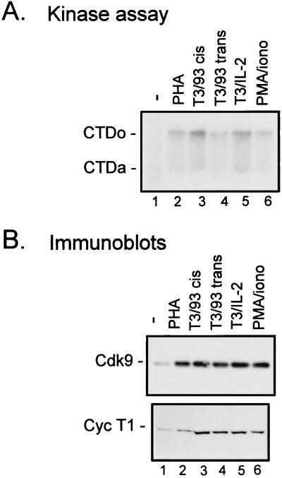
FIG. 2.
Cdk9 and cyclin T1 protein levels are increased following activation of CD4+ T cells by a variety of activating stimuli. Primary CD4+ cells were cultured in media containing 10% FBS (−) (lanes 1) or with the addition of PHA (5 μg/ml) (lanes 2), antibodies against CD3 and CD28 (9.3) on the same bead complex (T3/93 cis) (lanes 3) or on separate beads (T3/93 trans; beads used at a ratio of 1 bead/cell) (lanes 4), IL-2 (100 U/ml) and antibody against CD3 (T3/IL-2) (lanes 5), or PMA (1.2 ng/ml) and ionomycin (0.08 μg/ml) (PMA/iono) (lanes 6). After 3 days, cells were lysed and equal amounts of protein were assayed for TAK activity by a kinase assay with recombinant CTD as a substrate (A) or for protein levels of Cdk9 or cyclin T1 (Cyc T1) by immunoblotting (B). CTDo, hyperphosphorylated form of the CTD; CTDa, underphosphorylated form of the CTD.

