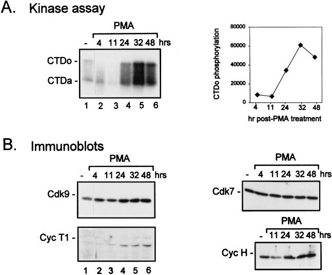
FIG. 6.
Time course of induction of cyclin T1 protein levels following PMA treatment of HL-60 cells. HL-60 cells were cultured in media containing 10% FBS (−) or with the addition of PMA (0.3 ng/ml) for the indicated times. Cell lysates were prepared, and equal amounts of protein were assayed for TAK activity by a kinase assay with recombinant CTD as a substrate (A) or for protein levels of Cdk9 or cyclin T1 (Cyc T1) by immunoblotting (B). Quantitation of the TAK assay was performed by measurement of CTDo phosphorylation as determined by PhosphorImager scanning. CTDo, hyperphosphorylated form of the CTD; CTDa, underphosphorylated form of the CTD; hr and hrs, hours.

