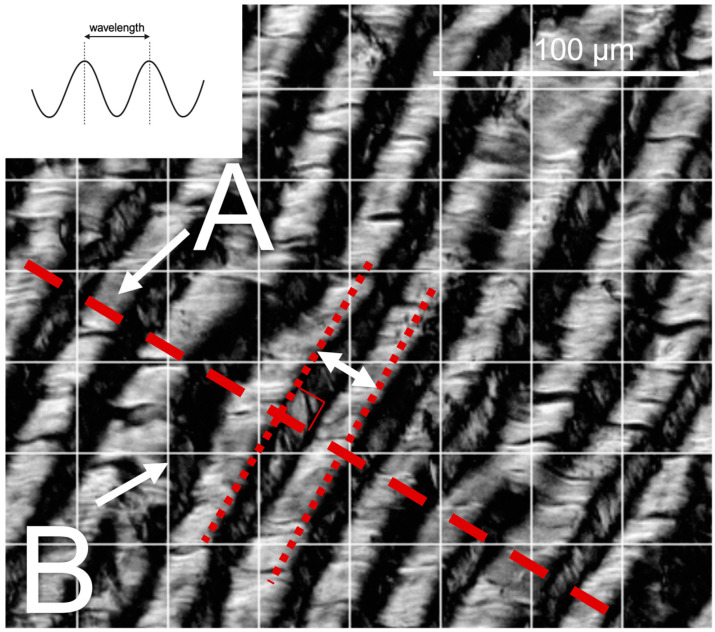
Figure 1.
Methods used for collagen crimp measurement. The inset schematic depicts wavelength measured as the distance between adjacent “peaks” in the crimp waveform. Representative polarized light microscopy image shows crimp length measurements along a line normal to the crimp direction (A). Crimped area measurements were taken at each intersection of gridlines (B), as the ratio of crimped-to–total-grid-points (grid = 0.01 mm2/square) to calculate the area percentage of tissue occupied by crimp.

