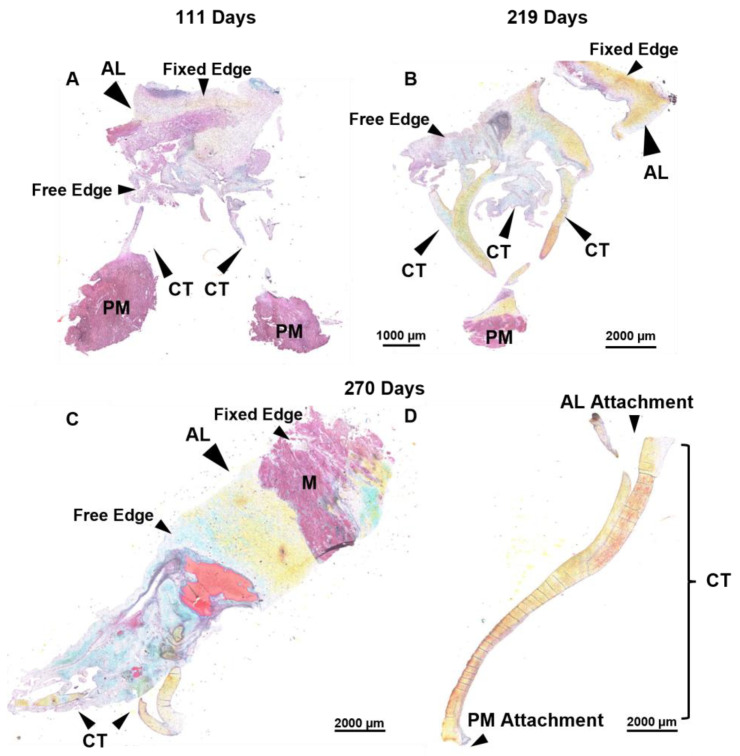
Figure 7.
Throughout the second trimester to full term, the anterior leaflet and chordae tendineae are comprised predominantly of collagen. (A–D) Representative images of extracellular matrix staining (Movat Pentachrome) from early second trimester (111 days gestation), third trimester (219 days gestation), and full term (270 days). Black arrows denote the anterior leaflet (AL) and chordae tendineae (CT). Papillary muscle (PM) and myocardium (M) are labeled. Note that these are transverse longitudinal sections, therefore the fixed edge and the free edge are labeled for orientation. For the strut chordae in (D), anterior leaflet attachment and papillary muscle attachment are labelled for orientation. Collagen is stained yellow-orange, elastic fibers dark purple, muscle tissue and blood cells red, glycosaminoglycans blue-green, and cell nuclei dark red-purple. Scale bar varies per image.

