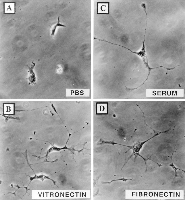
FIG. 5.
Vitronectin and fibronectin permit the formation of VV-induced cellular projections. BS-C-1 cells were infected with VV and were transferred to glass coverslips pretreated with PBS (A), 10 μg of vitronectin/ml (B), FBS (C), or 10 μg of fibronectin/ml (D). The morphology of cells was recorded at 18 hpi after fixation for 1 h in 0.1% crystal violet.

