Figure 2.
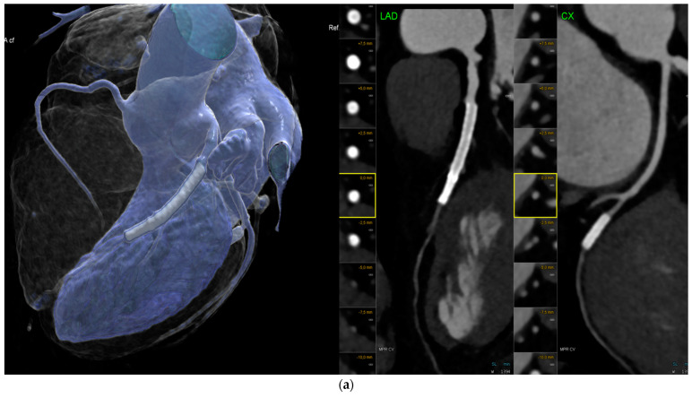
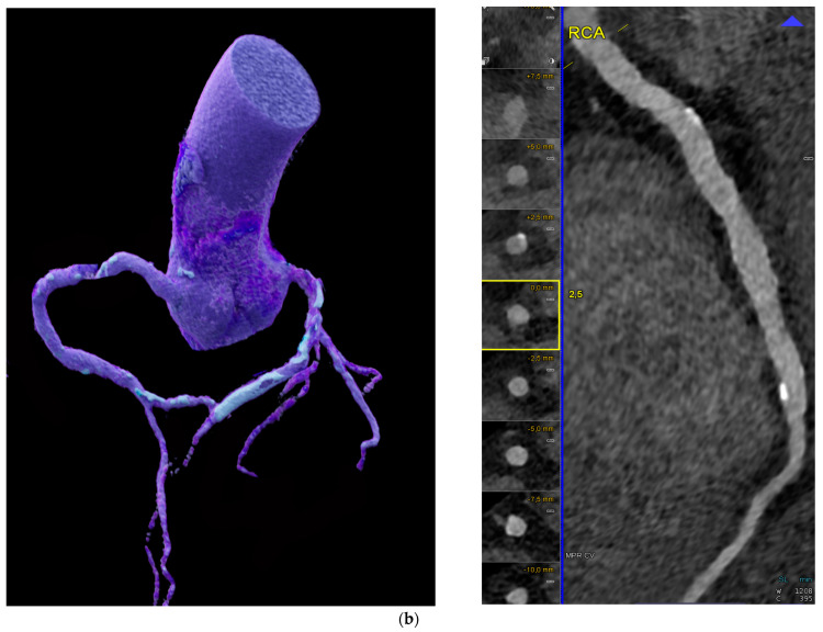
(a). Imaging of coronary stents by conventional energy-integrating detector (EID)-CT: Multiple left anterior descending (LAD) coronary stents with inadequate in-stent lumen visualization of the distal LAD and circumflex artery (CX) stent due to artifacts. Left: volume-rendering technique (VRT) and right: curved multiplanar reformations (cMRP) of the LAD and CX. (b). Photon-counting detector CT (PCD-CT): Curved multiplanar reformation (cMRP) (left) shows calcified right coronary artery (RCA) in a 76-year-old male patient with a high calcium load, but reduced blooming artefacts with less than 50% stenosis. Stent in the mid-left anterior descending (LAD) proximal coronary artery (VRT, right panel) and heavy vessel wall calcification. VRT = volume-rendering technique.


