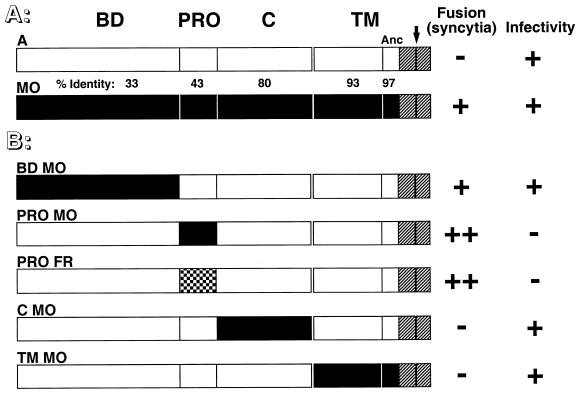
FIG. 1.
Schematic representation of envelope chimeras and their fusion properties. White and black boxes represent domains derived from amphotropic MLV-4070A envelope (A) and ecotropic Moloney-MLV (MO) or Fr-MLV (FR) envelope, respectively. The intracytoplasmic sequences, identical for both MLV classes, are shown as hatched boxes. (A) Domain organization of parental envelope. BD, amino-terminal receptor binding domain; PRO, proline-rich region; C, SU carboxy-terminal domain; TM, transmembrane subunit. Separation between ectodomain and anchor domain (Anc) of the TM subunit is indicated by the thin vertical black bar. The percentage of identical amino acids between each domain is indicated. The black arrow over the 4070A-MLV TM subunit marks the location of a premature stop codon introduced immediately before the R peptide to generate the cell-to-cell fusogenic ARless amphotropic envelope. (B) Chimeric envelope in which single domains were swapped. A summary of fusion and infection properties is shown to the right of the schematic representations. Cell-to-cell fusion activity was determined after transfection of the corresponding envelope expression vector in TELac2 cells and cocultivation with XC or XC-A-ST cells and is indicated as follows: −, absence of syncytia; +, presence of syncytia in XC cells; ++, presence of syncytia for chimeras with the amphotropic BD in both XC and X-A-ST cells (see detailed results in Table 1). Infectivity was tested by using supernatants harvested from stably transfected TELCeB6 packaging cells on XC cells and is indicated as follows: −, titers of less than 102 IU/ml; +, titers of greater than 106 IU/ml.

