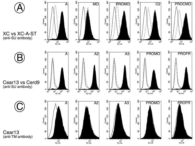
FIG. 5.
Binding assays of mutant envelopes with enhanced fusion activity. Binding assays were performed with supernatants of TELCeB6 cells transfected with the indicated envelope expression vectors depicted in Fig. 1, 4, and 7 on PiT-2-expressing XC cells and Cear13 cells (black area), on PiT-2-interfering XC-A-ST cells (broken lines, panel A) or on PiT-2-negative Cerd9 cells (broken lines, panel B), as indicated. The background fluorescence was provided by incubating XC or Cear13 cells with supernatant of nontransfected TELCeB6 cells (solid lines). The background fluorescence on XC-A-ST cells and on Cerd9 cells (not shown) was the same as that on XC cells and Cear13 cells, respectively. Incubated cells were stained with the indicated antienvelope antibodies. The envelope glycoprotein content of the different samples was normalized by immunoblotting of viral supernatant (A and B) or viral pellet (C).

