Figure 1.
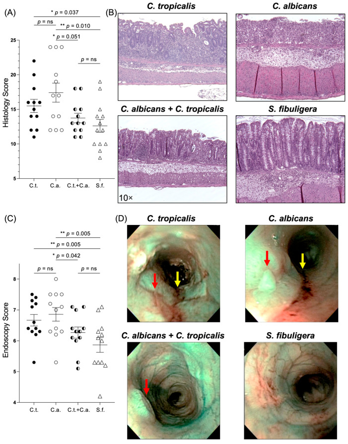
Candida (C.) tropicalis inoculation decreased susceptibility to chemical-induced colitis in C. albicans-challenged C57BL/6 mice. (A) Histological analysis showed decreased colonic inflammation in co-inoculated mice compared to mice challenged with C. albicans alone (unpaired t-test, 13.75 ± 0.69 vs. 17.50 ± 1.36; p < 0.05; N = 12/group) and increased colonic inflammation in C. albicans-challenged mice and C. tropicalis-challenged mice compared to control group (17.50 ± 1.36 vs. 12.62 ± 0.93; p < 0.02) (15.50 ± 0.95 vs. 12.62 ± 0.93; p < 0.05). No statistical differences were found between co-inoculated mice and control group (13.75 ± 0.69 vs. 12.62 ± 0.93; p = ns) or between co-inoculated mice and C. tropicalis-inoculated group (13.75 ± 0.69 vs. 15.50 ± 0.95; p = ns). (B) Representative colonic histopathological sections of C. albicans- and C. tropicalis-inoculated mice show the presence of ulcers, active cryptitis, increased inflammatory cells in the lamina propria, and thicker intestinal mucosa compared to co-inoculated mice and control group, showing minimal inflammatory cells and mild active cryptitis. (C) Colonoscopic evaluation showed increased colitis in distal colon of mice challenged with C. albicans alone compared to co-inoculated mice (6.86 ± 0.22 vs. 6.27 ± 0.18; p < 0.05) and control group (6.86 ± 0.22 vs. 5.87 ± 0.24; p < 0.02). No statistical differences were found between co-inoculated mice and control group (6.27 ± 0.18 vs. 5.86 ± 0.24; p = ns) or between co-inoculated mice and C. tropicalis-inoculated group (6.27 ± 0.18 vs. 6.67 ± 0.19; p = ns). (D) Narrow-band imaging endoscopic pictures of distal colon showed higher presence of ulcers (red arrows) and colorectal bleeding (yellow arrows) in C. albicans-inoculated and C. tropicalis-inoculated mice compared to co-inoculated mice and control group. Data are expressed as mean ± SEM; * p < 0.05, ** p < 0.02.

