Figure 5.
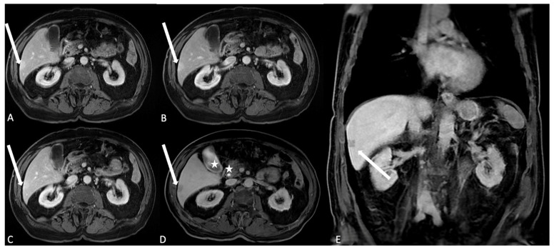
T1 LAVA axial images after HSCA (gadobenate dimeglumine) intravenous administration. (A) The arterial phase. Arterial structures and hypervascular lesions are evidenced: in the liver segment 6, it is possible to identify a subcapsular hyperintense area as indication of a hypervascular behavior (arrow); (B) in the venous phase, the liver parenchyma reach the best enhancement and the hypervascular area in segment 6 shows persistent enhancement (arrow); (C) the delayed/equilibrium phase allows representation of interstitial and extracellular spaces enhancement. The subcapsular lesion is quite completely homogeneous to the liver parenchyma arrow, suggesting the angiomatous nature of the lesion. (D,E) Axial and coronal images after HSCA (Multihance) intravenous administration of the same patient acquired in the delayed hepatobiliary phase show the opacification of the gallbladder lumen and the choledocic duct (stars); the subcapsular lesion is also identified as the hypointense area (arrows), confirming the vascular nature of the angioma and the lack of hepatocyte uptake and biliary excretion.

