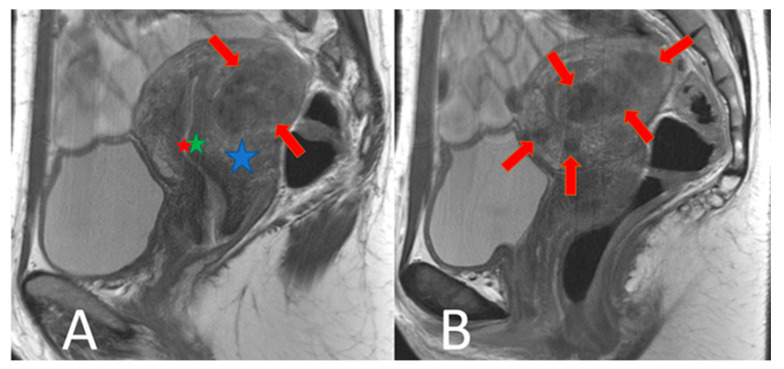
Figure 2.
Focal adenomyosis: T2w sagittal images. (A): The uterus is enlarged by multiple T2 non-homogeneously hypointense nodules of different sizes in the outer myometrium (red arrows). The endometrium (red star) and the inner myometrium (green star) are outlined. The outer myometrium (blue star) is partly visible. (B) shows another analogous example.

