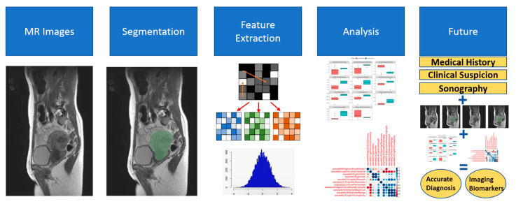
Figure 4.
Schematic representation of segmentation, feature extraction, analysis, and future potential of radiomics. This figure illustrates the process of how the radiomics features were derived from T2w MRI and especially illustrates how the image segmentation and feature extraction were performed. Additionally, the future potential of using radiomics in combination with clinical data is highlighted.

