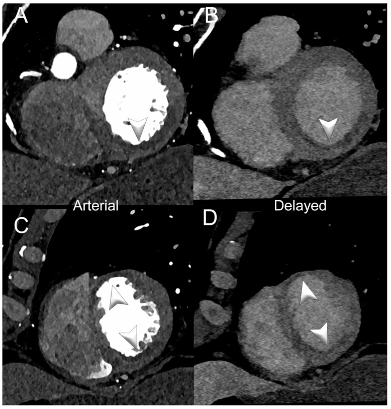
Figure 3.
Spectral cardiac/coronary PCCT example of sub-acute LV ischemia. The figure shows a patient with a recent acute myocardial infarction in the territory of the right coronary artery and of the left anterior descending coronary artery. Panels (A,B) show a short-axis view of the base of the left ventricle (LV) in arterial/angiographic phase (A) and delayed phase (B); panels (C,D) show the same information in the apical section of the LV. Arrowheads indicate the multiple LV segments with hypoperfusion (early and late). The delayed phase was performed with spectral acquisition and monochromatic+ 40 KeV reconstruction. The scan was performed on a commercial whole-body Dual Source Photon-Counting CT scanner (NAEOTOM Alpha, Siemens Healthineers), with 0.2/0.4 mm slice thickness, 0.1/0.2 mm reconstruction increment, FOV 140–160 mm, resolution matrix of 512 × 512/1024 × 1024 pixels on the source axial reconstructions with a kernel filtering of Bv48-60 (vascular kernel medium-sharp) and with maximum intensity of Quantum Iterative Reconstruction (QIR 4); the scan is performed with retrospective ECG gating with tube current modulation. The displayed spatial resolution is 0.1/0.20 mm. Abbreviations: PCCT = Photon-Counting CT; LV = left ventricle; ECG = electrocardiographic.

