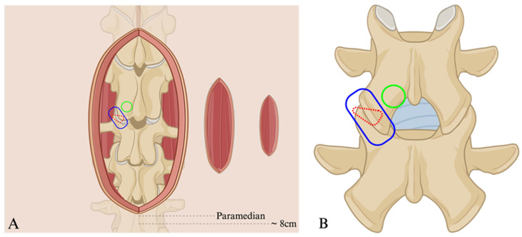
Figure 2.
Animated depiction of various access trajectories at the level of the skin (A) and target vertebral level (B). Wide midline incision (A) representing the exposure for the PLIF and open TLIF. More laterally, paramedian is a representation of the skin incision for the MIS-TLIF and the transfacet LIF; most laterally is the representative incision for the Trans-Kambin’s Triangle TLIF at approximately 8 cm from midline. The green circle highlights (A,B) the point of access osteotomy for the TLIF and blue encircles the facet joint, which represents the transfacet corridor. Finally, the red dashed triangle is a depiction of where Kambin’s Triangle would lie from a lateral viewpoint—dashes indicate that the triangle is not viewable from this posterior angle. Figure created using BioRender.com.

