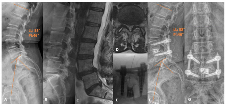
Figure 3.
(A) Preoperative standing X-ray demonstrating the L4-5 grade 1 spondylolisthesis, with (B) flexion films demonstrating dynamic instability. (C) Sagittal and (D) axial T2-weighted MRI slices revealing the advanced central and bilateral recess stenosis at the L4-5 level. (E) Intraoperative fluoroscopic image following cage placement with a guidewire within the disc space. (F) Postoperative lateral and (G) AP X-rays supporting a satisfactory appearance of the construct. LL: Lumbar lordosis. PI: Pelvic incidence.

