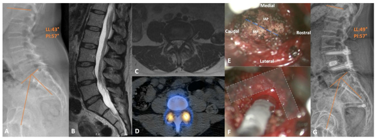
Figure 4.
(A) Preoperative standing lateral X-ray demonstrating the L4-5 grade 1 spondylolisthesis. (B) Sagittal and (C) axial T2-weighted MRI showing the mild central and severe lateral recess stenosis, respectively, with the latter image depicting the bilateral facet joint effusion. (D) Axial slice of the CT SPECT scan showing the isolated increase in radiotracer uptake at both L4-5 facet joints. (E) Intraoperative view of a right-sided transfacet approach with the joint line (dashed line), inferior articular process (IAP), and superior articular process (SAP) illustrated. (F) The same approach after completing the required facetectomy and discectomy to allow adequate room for cage trials; bony boundaries protecting the neural structures (L shape). (G) Postoperative lateral X-ray with adequate reduction in the slip and improved lordosis.

