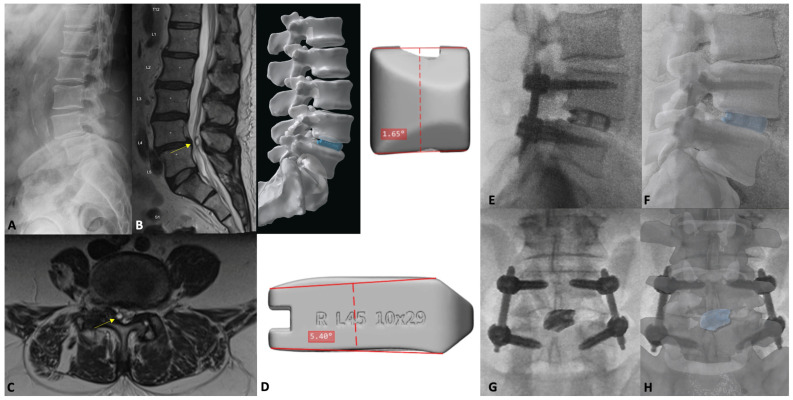
Figure 7.
Lateral flexion plain film (A) demonstrating the L4-5 spondylolisthesis and disc height loss. Sagittal (B) and axial (C) T2-weighted MRI depicting the L4-5 facet cyst (yellow arrow) and lateral recess stenosis. Preoperative 3D rendering (D) of the patient’s spine and proposed custom L4-5 interbody (Carlsmed aprevo, Carlsbad, CA, USA). Postoperative lateral (E) lumbar plain film with overlying custom cage rendering (F). Postoperative AP (G) plain film with overlying custom cage rendering (H).

