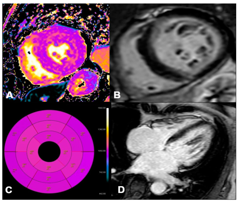
Figure 6.
AL 73-year-old patient diagnosed with endomyocardial biopsy. T1 Native Mapping (A) demonstrates a slight increase in value in all entire LV walls with subendocardial patterns. Circumferential subendocardial LGE of the anterior, antero-lateral, and infero-lateral LV wall in two-chamber short (B) and of the SIV to the apical segment in long axes (C). Bullseye map of T1 Native (D). LV, left ventricle; LGE, late gadolinium enhancement.

