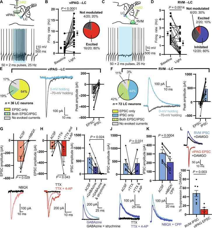
Fig. 6. Electrophysiological characterization of inputs from the vlPAG and RVM to the LC.
(A) Top: vlPAG ChR2 viral injection for LC slice electrophysiology. Bottom: Representative trace of tonic spiking during a 2-s blue LED stimulus (470 nm, 50 × 2-ms pulses, 25 Hz, 18 mW). (B) Left: Firing rate before and during the stimulus (n = 20 neurons; two-sided Wilcoxon matched-pairs signed rank test). Right: Categorization of neurons as “excited” (z score of spiking during versus before light > 2), “inhibited” (z score < −2), or “not modulated.” (C) Same as (A) for ChR2 expressed in the RVM. (D) Same as (B) for RVM terminal stimulation (n = 20 neurons; two-sided Wilcoxon matched-pairs signed rank test). (E) Left: Proportion of LC neurons with oEPSCs and oIPSCs during vlPAG terminal stimulation (1 × 5-ms pulse, 18 mW). Middle: Representative example of an oEPSC and oIPSC in a single LC neuron. Right: Peak amplitude of oEPSCs and oIPSCs (n = 12 neurons). (F) Same as (E) for RVM terminal stimulation (n = 12 neurons). (G to K) Top: Summary bar graphs of oEPSC/IPSC amplitude. Bottom: Representative examples. (G) NBQX effect on vlPAG oEPSC amplitude (two-sided paired t test: t = 3.950, n = 4 pairs). (H) TTX and 4-AP effect on vlPAG oEPSC amplitude (two-sided paired t test: t = 2.926, n = 5 pairs). (I) Effect of GABAzine and strychnine on RVM oIPSC (repeated-measures one-way ANOVA with Dunnett’s multiple comparisons test, P = 0.013, F1.087,4.348 = 16.32, n = 5 cells). (J) Same as (H) for RVM oIPSCs (two-sided Wilcoxon matched-pairs signed rank test, n = 6 pairs). (K) Effect of NBQX and CPP on RVM oIPSC amplitude (two-sided paired t test: t = 6.976, n = 7 pairs). (L) Top: Examples of RVM oIPSCs (blue) and vlPAG oEPSCs (red) after bath application of DAMGO (1 μM; black). Bottom: Opioid sensitivity reported as % suppression of amplitude (two-sided unpaired t test: t = 3.785, n = 8 RVM oIPSC, n = 5 vlPAG oEPSC). All summary data reported as means ± SEM.

