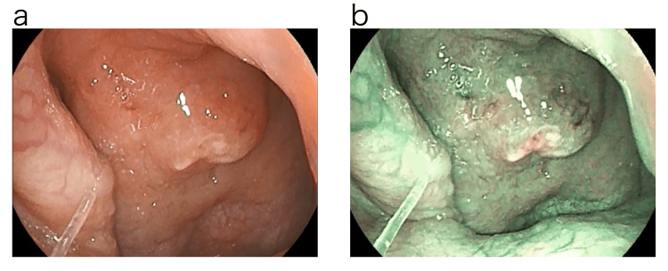
Figure 3. Endoscopic findings of the epipharynx before treatment.
Examination using normal light revealed swelling of the epipharyngeal mucosa centered on the Thornwald site and an accumulation of an abscess. Examination using optical enhancement mode 1, which uses band-limited light, revealed reddish-brown or dark-brown findings on the mucosal surface that may indicate internal hemorrhage. The deep mucosa showed a greenish color, which may indicate deep vascular dilatation or congestion.
a: Normal light.
b: Optical Enhancement Mode 1.

