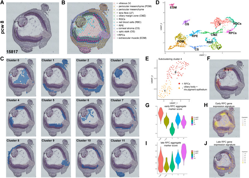
Fig. 2. ST analysis of 8 PCW human eye sections reveals the location of early RPCs in the CMZ.
A Representative histological staining of the 8 PCW fresh frozen human eye section. Four sections from the same eye sample were processed for ST analyses. B, C Spatial localisation of the 12 clusters identified from the ST analysis. Highly expressed markers for each cluster are shown in Supplementary Data 3. D UMAP of spatial transcriptomics scRNA-Seq data. E Subclustering of ciliary margin zone (cluster 4) reveals the presence of two subclusters namely RPCs and ciliary body, and iris pigmented epithelial cells: their spatial localisation is shown in panel (F). G and (I) Expression violin plots showing the highest aggregate expression scores for early RPCs in the peripheral retina (CMZ) and late RPCs in the central retina respectively, compared to all other retinal clusters identified in the ST analysis. H and (J) Early and late RPC gene expression signatures superimposed on the spatial image of 8 PCW human eye.

