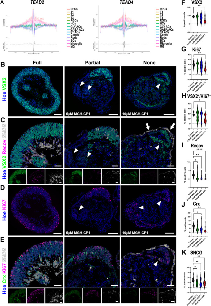
Fig. 7. TEAD binding plays a significant role in RPC proliferation.
A Footprinting analysis of TEAD2 and TEAD4 showing a significant enrichment in RPCs. Additional abbreviations to those mentioned in the main text: Gly ACs- glycinergic amacrine cells, GABA ACs – gabaergic amacrine cells, ST ACs – starburst amacrine cells, HCs – horizontal cells, MG- Muller glia cells. B–K Quantitative immunofluorescence analyses for the presence of VSX2+ RPCs (B, F), Ki67+ proliferating cells (D, E, G), VSX2+Ki67+ (H), SCNG+ RGCs (C, E, K), and Recoverin+ (C, I) and CRX+ photoreceptor precursors (E, J) reveal loss of RPCs, disturbed retinal lamination, and attenuation of photoreceptor and RGCs specification. Bottom panel insets at (C) and (E) panels show individual antibody and nuclear staining. White arrowheads show the presence of rosettes comprised of RPCs or Ki67+ proliferating cells. Scale bars 100 µM for (B–D) and 50 µM for (C–E) and bottom inset panels. F–K Data presented as median and quartiles. 9 – 39 retinal organoids per condition were used as documented in the Source data file. One-way ANOVA (F–H) or Kruskal-Wallis (I–K) with Dunnett’s multiple comparisons test (*p < 0.05; **p < 0.01, ***p < 0.001). Source data are provided as a Source Data file.

