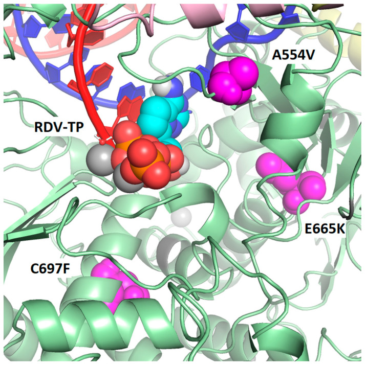
Figure 3.
Map of observed post-baseline amino acid substitutions closest to the active site of Nsp12 and on cryo-EM structure of the SARS-CoV-2 polymerase complex with pre-incorporated RDV-TP. None of the observed post-baseline substitutions is in direct contact with RDV-TP or the RNA primer or template strands. However, three of the substitutions are located within 20 Å of the pre-incorporated RDV-TP (as measured from Cα to C1′). A554V is 13.9 Å, C697F is 16.9 Å, and E665K is 18.0 Å. The Nsp12 protein is green with the locations of the substitutions shown in magenta. The template RNA strand is shown in blue and nascent RNA strand in red. Pink is Nsp7, and yellow is Nsp8 (two subunits).

