Figure 9.
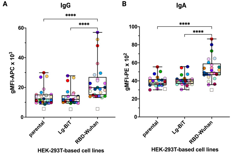
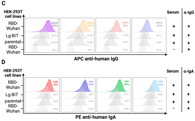
The FCCA may detect low levels of pre-existing cross-reactive anti-RBD antibodies in non-SARS-CoV-2-exposed individuals (Group A). Shown in (A,B) are pre-existing IgG and IgA antibodies binding to the SARS-CoV-2 RBD-Wuhan-Hu-1 in the SARS-CoV-2 non-exposed group A of individuals (n = 25). Y-axes show raw gMFI values. The reactivities with the parental HEK-293T cell line as well as against the transduction control HEK-293T-Lg-BiT stable cell line are shown as controls. Each dot represents the reactivity of a single serum sample (dilution 1:100) with the three different cell lines (parental, Lg-BIT, and RBD-Wuhan-Hu-1). The open squares show the reactivity (gMFI) of the respective secondary antibodies with the three different cell lines in the absence of prior incubation with serum. In the plot, the boxes indicate the upper and lower quartiles, with the horizontal line showing the median, with whiskers representing minimum and maximum values. ****, p < 0.0001 as determined by the Friedman test with Dunn’s multiple comparison correction. (C,D) Stacked histograms of the IgG or IgA reactivity in (A,B), respectively, of the four sera upon incubation with the RBD-Wuhan-Hu-1 transfectants compared to control transfectants (Lg-BiT) or parental HEK-293T cells (similar color code as in A,B). Sera were selected on the basis of high and low binding characteristics. In each histogram, the gMFI values are indicated and the percent positive values resulting from the comparison of serum reactivity between RBD-Wuhan-Hu-1 and Lg-BiT control transfectants are shown in parentheses. In the bottom-line histograms of each panel, the reactivity of the secondary antibody alone, in the absence of serum, to RBD-Wuhan-Hu-1 transfectants is shown.


