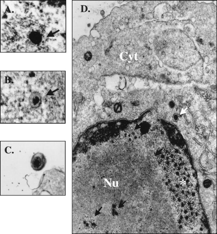
FIG. 4.
Visualization of KSHV in BKS-1 by TEM. (A and B) High (×82,400) magnification of a typical nucleocapsid with an electron-dense core seen in the nuclei of BKS-1 (A) and BCBL-1 (B) cells. (C) An enveloped particle morphologically mature in the extracellular space of a BKS-1 cell (magnification, ×25,900). (D) General view of a TPA-treated BCBL-1 cell. Numerous empty capsids or capsids with an electron-dense core (dark arrows) are present within the nucleus (Nu) of the cell. Electron-opaque bodies showing sites of assembly of capsids within the nucleus (*) and a cytoplasmic (Cyt) vesicle containing an enveloped mature particle (arrow) can also be seen (magnification, ×25,900).

