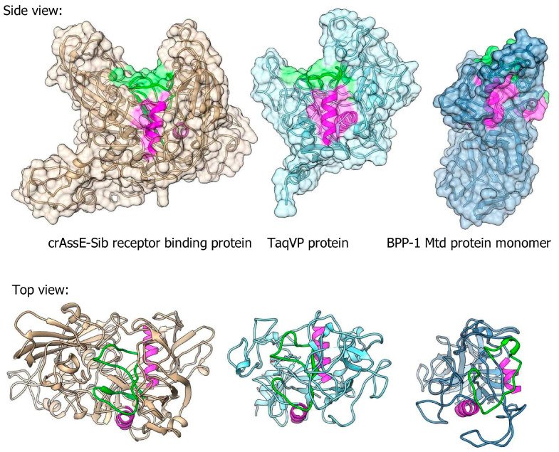
Figure 10.
Comparison between 3D model of crAssE-Sib receptor binding protein (RBP) and experimental structures of TaqVP protein from Thermus aquaticus (pdb id 5VF4) and Mtd protein of the BPP-1 phage (pdb id 1YU0). VR regions are in green; magenta alpha helices show similar orientation of the molecules. All molecules are on the same scale.

