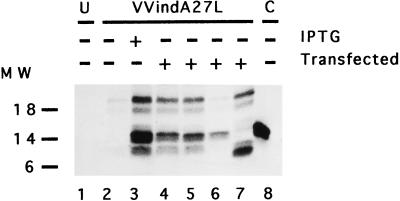
FIG. 8.
In vivo assay to measure 14-kDa–21-kDa protein-protein interactions. BSC40 cells grown in 12-well plates were infected (10 PFU/cell) with VVindA27L and transfected with plasmid vectors (10 μg/well) expressing different mutant forms of the 14-kDa protein. The cells were labeled with [35S]methionine-cysteine from 6 to 24 h p.i., and cell extracts immunoprecipitated with MAbC3 were analyzed by SDS-PAGE and autoradiography, as described in Materials and Methods. Lane 1, uninfected cells (U); lanes 2 to 7, cells infected with VVindA27L; lane 8, cells infected with VVindA17L (control [C]). Cells in lanes 4 to 7 were transfected with pHLZ-14K-wt, pHLZ-14K-A, pHLZ-14K-A-L89A, and pHLZ-14K-A-Δ29, respectively. Molecular weight (MW) markers are on the left. Densitometric analyses revealed the following ratios for the 14-kDa/21-kDa proteins normalized after subtracting the value in lane 2 (negative control): 1.49 (lane 3), 1.36 (lane 4), 1.3 (lane 5), 162.9 (lane 6), and 1.2 (lane 7).

