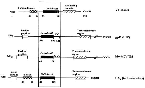
FIG. 9.
Schematic comparison of the VV 14-kDa protein, HIV gp41, Mo-MLV TM, and influenza HA2 structures. The four proteins form three-stranded coiled-coil structures involving a central α-helix. For all of them, the hydrophobic fusion peptide would be immediately amino terminal to the oligomerization domain, although for the VV 14-kDa protein the peptide implicated in this process has not been defined yet (22). For the 14-kDa protein an anchoring domain is indicated instead of a transmembrane region of the C terminus. Except for the influenza HA2, these fusion proteins have cysteine residues at the end of the coiled-coil region.

