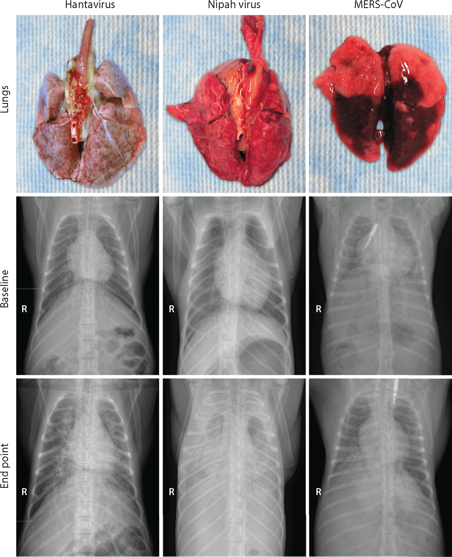
Figure 3.

Gross pathological and radiographic changes in the lungs of nonhuman primates experimentally infected with a hantavirus, Nipah virus, or Middle East respiratory syndrome coronavirus (MERS-CoV). Lungs were collected from a Sin Nombre virus inoculated rhesus macaque at 15 days postinoculation (dpi); from a Nipah virus, strain Malaysia, inoculated African green monkey at 4 dpi; and from a MERS-CoV inoculated common marmoset at 6 dpi. Radiographs collected from these same animals are displayed. The top radiographs represent baseline; the bottom radiographs were taken at the endpoint of the experiment. The radiopaque objects in the two MERS-CoV radiographs are subcutaneously injected temperature transponders.
