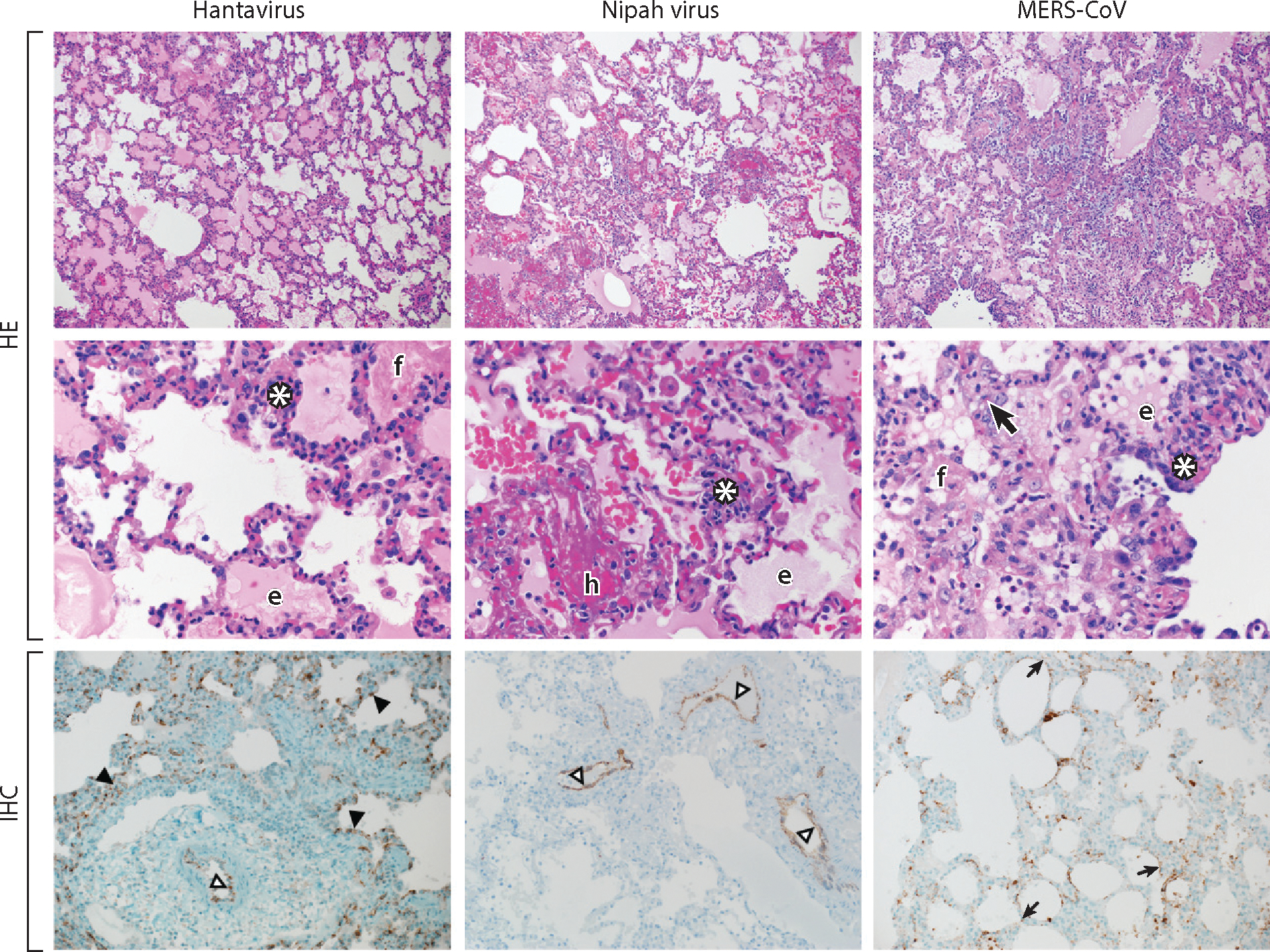
Figure 4.

Histopathological changes in the lungs of nonhuman primates experimentally infected with a hantavirus, Nipah virus, or Middle East respiratory syndrome coronavirus (MERS-CoV). Lungs were collected from a Sin Nombre virus inoculated rhesus macaque; from a Nipah virus, strain Malaysia, inoculated African green monkey; and from a MERS-CoV inoculated common marmoset. Lung tissue was stained with hematoxylin eosin (HE) or a virus-specific antibody (IHC). Viral antigen is shown as brown-red staining.
Abbreviations and symbols: white asterisks, inflammatory cells; f, fibrin; e, edema; h, hemorrhage; bold black arrow, type II pneumocyte hyperplasia; black arrowheads, alveolar capillary endothelium; open arrowheads, arterial endothelium; small black arrows, alveolar pneumocytes. Magnification: top panels, 100×; middle panels, 400×; bottom panels, 200×.
