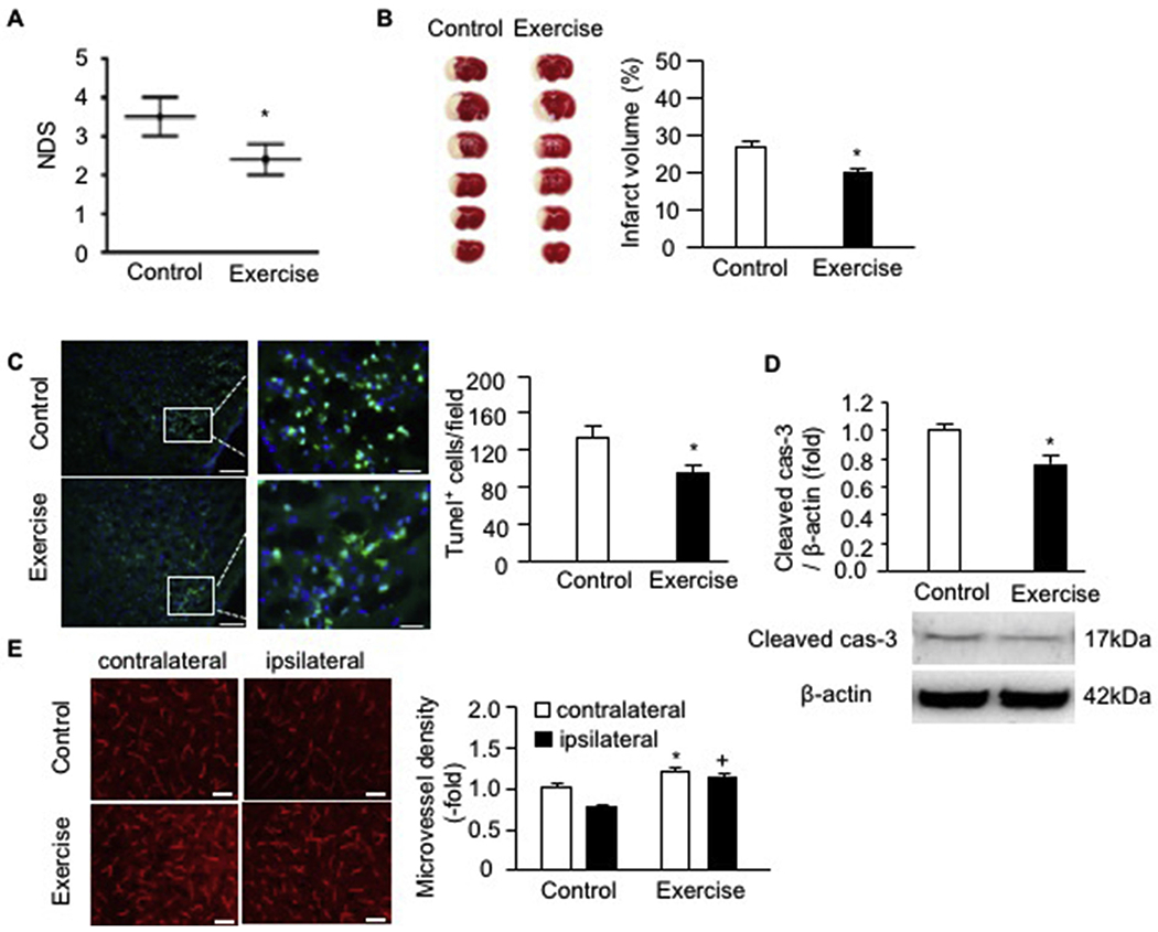
Fig. 1.

Exercised mice had reduced NDS and infarct volume, decreased cell apoptosis as well as a higher microvessel density on day 2 post-MCAO surgery. A, NDS; B, representative TTC images and summarized data showing the infarct volume of exercised and control mice; C, representative Tunel staining images and summarized data showing cell apoptosis in the peri-infarct area of exercised and control mice; Green: Tunel labeling; Blue: DAPI staining; scale bar in left panels: 100 um; scale bar in right panels (enlarged images of the box): 25 um; D, cleaved caspase-3 expression in ipsilateral brain of exercised and control mice; E, representative images and summarized data showing the microvessel density in the contralateral and ipsilateral brain; scale bar: 50 um. * p < .05, vs. control; + p < .05, vs. ipsilateral. Data are expressed as mean ± SEM. N = 11/group. (For interpretation of the references to colour in this figure legend, the reader is referred to the web version of this article.)
