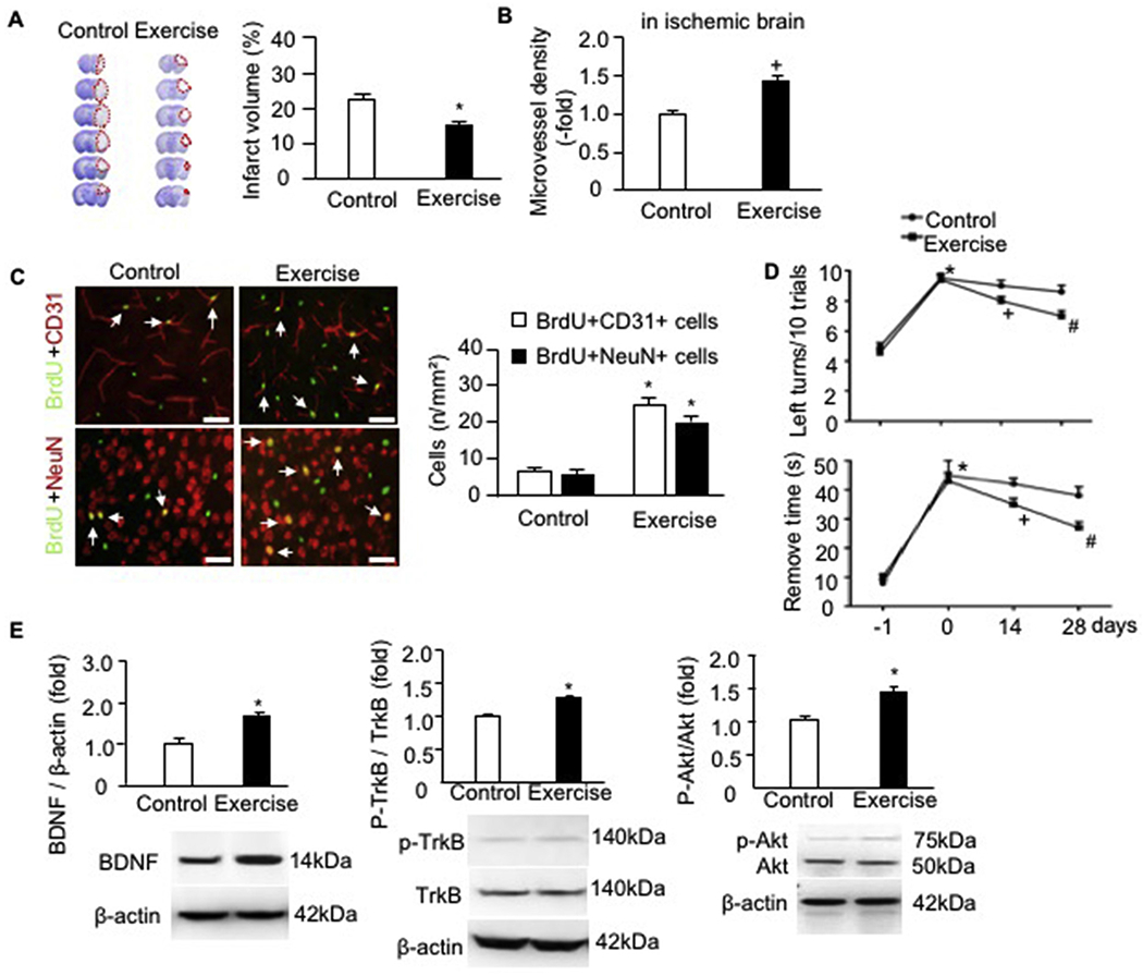
Fig. 4.

Exercised mice had alleviated infarct volume, increased microvessel density, angiogenesis and neurogenesis, improved sensorimotor functions as well as upregulated the protein expressions of BDNF/TrkB/Akt signaling pathway proteins in the ischemic brain of exercised mice day 28 post-MCAO surgery. A, representative CV images and summarized data showing the infarct volume of exercised and control mice; Red dash line indicate the infarct area in each brain slide; * p < .05, vs. control. B, microvessel density; C, representative immunofluorescence images and statistical data showing the angiogenesis and neurogenesis. Red: CD31 or NeuN; Green: BrdU; Double positive cells were labeled by arrows. Scale bar: 50 um. * p < .05, vs. control. D, corner test and adhesive remove test analyses. E, the protein levels of BDNF, p-TrkB/TrkB and p-Akt/Akt. * p < .05, vs. day −1, + p < .05, vs. day 0, # p < .05, vs. day 14. Data are expressed as mean ± SEM. N = 11/group. (For interpretation of the references to colour in this figure legend, the reader is referred to the web version of this article.)
