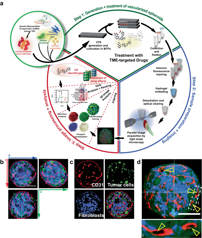
Fig. 1. Generation, treatment, preparation, and evaluation of vascularized tumor spheroids (VTSs).
a Schematic representation of the multistep process entailing pre-engineering of cell lines, generation of VTSs, treatment, sample preparation/staining, embedding, imaging, 3D-image processing, and image analysis, that enables the creation of datasets for a side-by-side comparison of complex structural effects triggered by TME-targeted drugs. b Preprocessed 3D-section image of a VTS generated with MDA-MB-435s cells (green, GFP-signal). Fibroblasts (blue, DsRed-signal). Incorporated ECs were stained for CD31 (red). c Compartmentalized representation of the xy-plane of the same VTS. The three cellular compartments are separated and processed. d Preprocessed section through a VTS generated with MDA-MB-468 cells. Yellow arrowheads show CD31+ PV structures with forming lumen. Scale bars = 100 µm.

