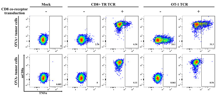
Figure 7. Functional testing of mouse-derived T-cell receptors (TCRs) requires co-transduction of mouse co-receptors.
Representative example of an experiment involving the functional testing of TCRs isolated from a transplantable mouse pancreatic ductal adenocarcinoma (PDAC) model expressing the chicken ovalbumin (OVA) neoantigen [18], approximately 50% of which were found directed against OVA [28]. In view of the proven reliability of our TCR-screening setup for human tumor–derived TCRs, we applied the same protocols for testing the anti-tumor reactivity of > 100 mouse PDAC tumor–derived TCRs. Shown in this figure is the reactivity of human CD8+ T222 cells transfected with a TCR derived from a tumor-infiltrating CD8+ T-cell clone, as compared to the OVA/SIINFEKL-specific TCR derived from the OT-1 TCR-transgenic mouse strain against IFNγ-pretreated OVA-expressing mouse PDAC tumor cells and the OVA-negative parental cells. The dot plots depict the expression of TNFα and mouse-CD8a+ in CD3+mTCRβ+ T-cells, thereby showing that both TCRs only mediate T-cell reactivity against the OVA-expressing tumor cells. Furthermore, the data demonstrate that reactivity by the strong OT-1-derived TCR is readily detected in the absence of co-transfection of the mouse CD8+ co-receptor, whereas detection of the reactivity of weaker TCRs is markedly facilitated by mouse-CD8 co-transfection. Notably, this does not significantly increase T-cell reactivity against the OVA-negative tumor cells.

