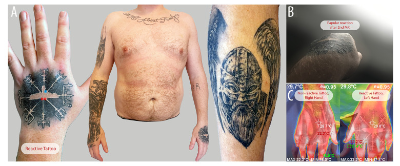
Figure 2.
(A) Overview of the patient’s tattoos. Left dorsal hand, reactive tattoo (early October 2022). Red circles indicate initial biopsy locations; the blue circle shows the second biopsy site post-MRI. The broader view depicts tattoos on the right arm (mid-2021 to early 2022), upper-chest lettering (2017), and a right leg tattoo (late 2021). (B) Reactive tattoo, a few days after the second MRI. The patient reported multiple papules in a follicular pattern within the black tattoo. The photo was taken by the patient in natural daylight. (C) IR thermography of the right versus left hand tattoo after the MRI test session. There was no significant thermal difference between the 2 hand tattoos.

