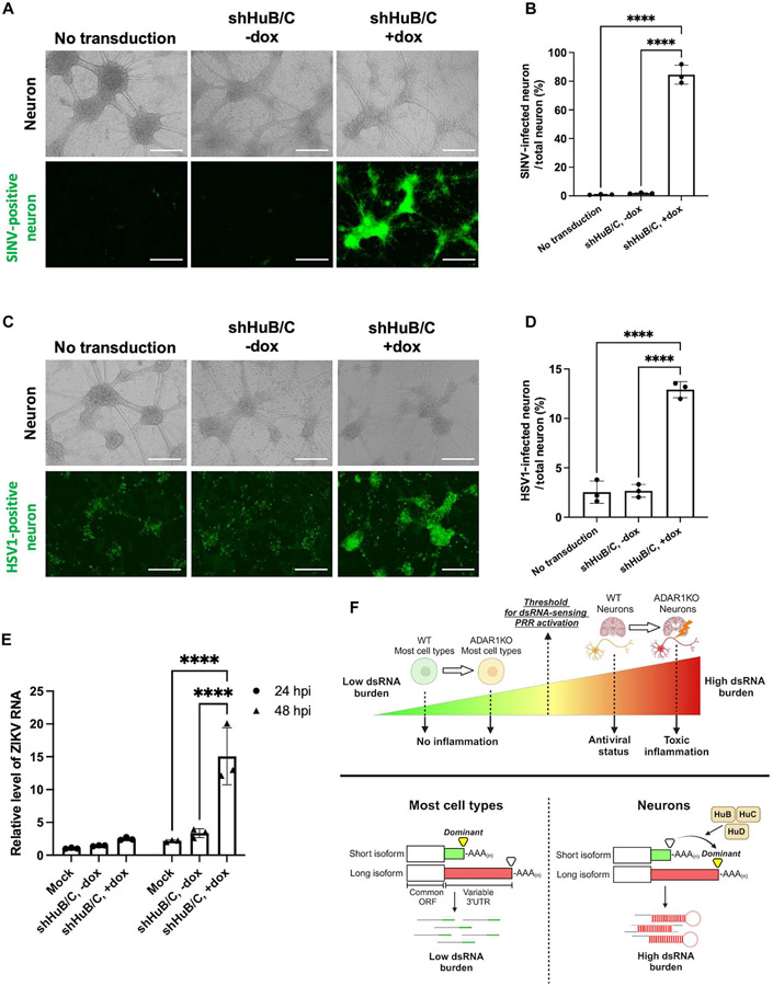
Fig. 7. HuB and HuC protect neurons from SINV, HSV-1, and ZIKV infection.
(A to E) Day 20 post-differentiation WT neurons were transduced with lentivirus containing doxycycline-inducible shRNA against HuB and HuC and then were infected with various viruses. (A and B) Neurons were infected with SINV (MOI = 0.1). (A) Fluorescent and brightfield images of neurons at 24 hours after infection. (B) Quantification of GFP-positive area in (A). (C and D) Neurons were infected with an HSV1 GFP reporter virus (MOI = 1). (C) Fluorescent and brightfield images of neurons at 24 hours after infection. (D) Quantification of GFP-positive area in (C). (E) Neurons were infected with ZIKV (MOI = 0.1), and infection was measured via qPCR of Zika RNA at both 24 (circle) and 48 (triangle) hours post-infection (hpi). (F) Summary graphic showing the main findings of this study. For Zika RNA qPCR, 18S RNA was used as a housekeeping gene. All quantified data shown are mean ± SD (n = 3 experimental replicates). Scale bars, 400 μm. (B and D) One-way ANOVA with Tukey corrected multiple comparisons. (E) Two-way ANOVA with Tukey corrected multiple comparisons, *P < 0.05, **P < 0.01, ***P < 0.001, and ****P < 0.0001.

