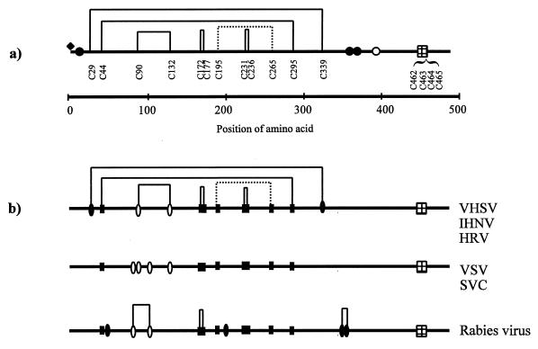
FIG. 4.
Rhabdovirus G proteins. (A) A schematic overview of the disulfide bonds and posttranslational modifications of VHSV G protein. Identified disulfides are shown as solid lines, and the predicted disulfide bond is shown as a dashed line. A filled diamond indicates the position of the O-glycan (Thr3). Filled circles indicate the positions of the characterized N-linked glycans (Asn10, Asn358, and Asn369), and the open circle indicates the position of a potential but not identified N-linked glycan (Asn418). The transmembrane regions are indicated by the gridded boxes. (B) Alignment of the G proteins from VHSV (GenBank accession no. ACX66134), IHNV (GenBank accession no. ACL40874), HRV (GenBank accession no. ACU24073), SVCV (GenBank accession no. ACU18101), VSV (GenBank accession no. ACM21421), and rabies virus, ERA strain (GenBank accession no. ACJ02293). Positions of conserved cysteine residues are marked by filled boxes, cysteines common to at least two groups are marked by open ovals, and unique cysteines are marked by filled ovals. Disulfide bonds in rabies virus are marked as reported by Dietzschold and coworkers (4).

