


Figure 3.
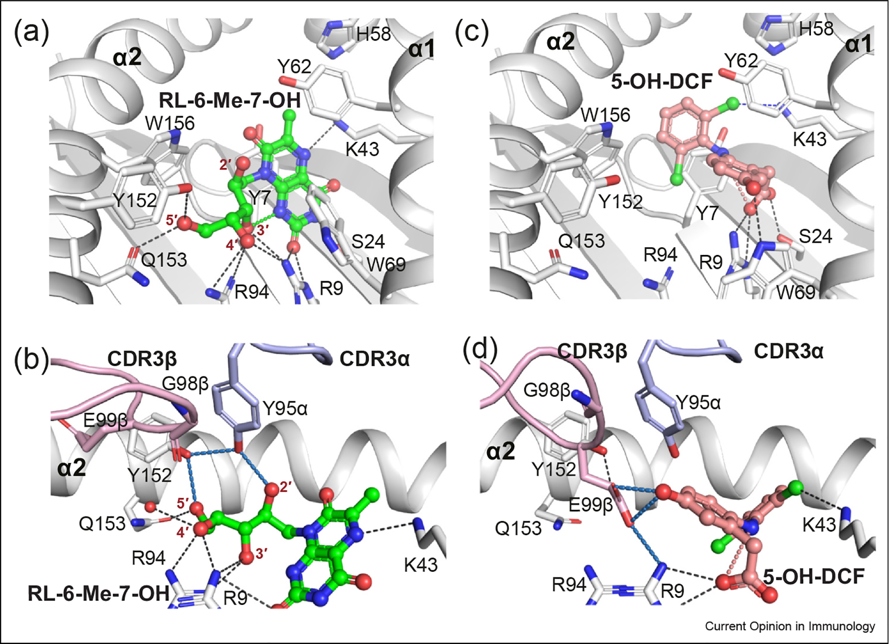
MR1 presentation of noncovalent MAIT agonists. The RL-6-Me-7-OH (a–b) and 5-OH-DCF (c–d) metabolites are shown as colored sticks in the MR1-binding pocket (upper panels) and their interactions with A-F7 MAIT TCR (bottom panels) (PDB: 4L4V and 5U72, respectively). RL-6-Me-7-OH and 5-OH-DCF ligands and their intramolecular H-bonds are colored green and salmon, respectively. Halogen bonds are colored in blue. H-bonds, CDR loops are colored as in Figure 2.